Flexible bronchoscopes have become indispensable tools in both diagnostic and therapeutic pulmonary medicine, offering clinicians unparalleled access to the tracheobronchial tree with minimal patient discomfort. Characterized by their slender, flexible shaft and fiberoptic or video-chip–based imaging, these instruments facilitate a broad spectrum of interventions—from simple airway inspection to complex bronchoscopic procedures. Below, we explore key aspects of flexible bronchoscope usage, illustrated in tabular form, and conclude with a concise summary.
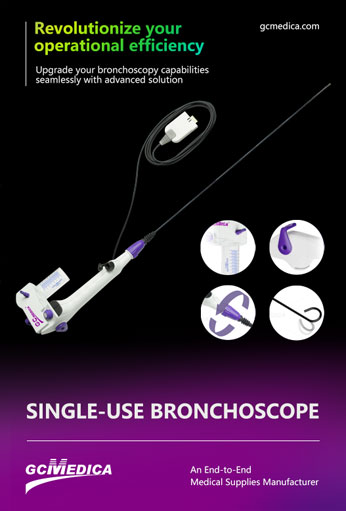
Flexible bronchoscopes typically consist of a control handle, insertion tube, light source, and working channel. The control handle allows precise tip deflection in multiple directions, enabling navigation through tortuous airway branches. The working channel accommodates biopsy forceps, cytology brushes, suction catheters, and other accessories, making it possible to perform tissue sampling, lavage, foreign-body removal, and stent placement. Video-chip technology has further enhanced image resolution and ergonomics, providing real‑time, high‑definition views on external monitors.
Patient preparation for flexible bronchoscopy involves topical anesthesia of the oropharynx (e.g., lidocaine spray), optional conscious sedation, and monitoring of vital signs. In intensive care or emergency settings, the procedure can be performed at the bedside, avoiding the risks of patient transport. In the operating room or bronchoscopy suite, general anesthesia is sometimes used for complex interventional cases. Throughout, careful attention to oxygenation and hemodynamic status is paramount.
The learning curve for flexible bronchoscopy is moderate: clinicians must develop hand–eye coordination and familiarity with airway anatomy under two‑dimensional visualization. Simulation training and supervised procedures accelerate competency acquisition. Once mastered, the flexible bronchoscope can be used across diverse clinical contexts, from routine outpatient diagnostics to advanced interventional pulmonology.
| Aspect | Details |
|---|---|
| Primary Uses | Diagnostic sampling (biopsy, bronchoalveolar lavage), airway inspection, foreign‑body removal |
| Imaging Technology | Fiberoptic vs. video‑chip; high‑definition monitors enhance visualization |
| Working Channel | Typically 2.0–2.8 mm diameter; accommodates forceps, brushes, suction, laser fibers |
| Patient Settings | Outpatient clinics, ICU/bedside, operating room; sedation or general anesthesia as indicated |
| Advantages | Minimally invasive, real‑time visualization, versatile accessory compatibility |
| Limitations | Narrow channel limits large‑bore tools; image quality lower than rigid scopes for stiffer applications |
| Maintenance | Requires high‑level disinfection or sterilization; prone to damage if mishandled |
| Cost Considerations | Capital investment in scope and processor; recurring costs for reprocessing and repairs |
Each aspect of flexible bronchoscope usage contributes to its role as a first‑line instrument in modern pulmonary care. The combination of maneuverability and accessory versatility makes it ideal for both routine and specialized procedures. However, limitations in channel size and potential for equipment wear necessitate careful case selection and diligent maintenance protocols.
Flexible bronchoscopes offer a balance of patient comfort and procedural capability that has reshaped respiratory interventions. Their flexible design allows safe navigation of complex airway anatomy, while a variety of accessories supports diagnostic and therapeutic goals. Although maintenance and procedural training requirements represent ongoing commitments, the clinical benefits—rapid bedside access, minimally invasive sampling, and expanded interventional options—make flexible bronchoscopy a cornerstone of pulmonary medicine.
| Single Use Flexible Bronchoscopy > |


 Français
 Español
 Products
 About Us


