Bronchoscopy is an essential procedure for visualizing and intervening in the tracheobronchial tree. Two primary modalities exist: flexible bronchoscopy (FB) and rigid bronchoscopy (RB). Each technique offers distinct advantages and limitations, and the choice depends on the clinical context, patient factors, and therapeutic goals.
Flexible bronchoscopy utilizes a thin, pliable fiberoptic or video‑chip scope that can navigate distal airways. Rigid bronchoscopy employs a straight, hollow metal tube and requires direct suspension of the patient’s head. Although both approaches share the goal of airway inspection, their instrumentation, anesthesia requirements, indications, and risk profiles differ substantially.
Detailed Comparison Table
| Feature | Flexible Bronchoscopy (FB) | Rigid Bronchoscopy (RB) |
|---|---|---|
| Instrumentation | 3–6 mm outer diameter, fiberoptic or video chip; multiple angulation points | 8–14 mm metal tube; straight, non‑angulating |
| Anesthesia/Sedation | Moderate sedation or local anesthesia; often performed awake or lightly sedated | General anesthesia with muscle relaxation; requires endotracheal control |
| Airway Control | Limited; relies on the patient’s spontaneous breathing | Secure; allows positive-pressure ventilation and airway suction |
| Visual Field | Good high‑resolution imaging of segmental bronchi | Excellent central airway view; limited distal reach |
| Working Channel | 1.2–2.8 mm channel; suitable for biopsies, brushes, and small instruments | Large channel (up to 7 mm); accommodates rigid forceps, stents, and debulking tools |
| Indications | Diagnostic sampling (biopsy, lavage), foreign body retrieval, airway inspection | Therapeutic interventions: tumor debulking, large foreign bodies, massive hemoptysis |
| Advantages | Minimally invasive, high patient tolerance, outpatient setting possible | Robust airway control, powerful suction, effective for large obstructions |
| Limitations | Less effective for massive bleeding or large rigid instruments | Requires OR setting, higher procedural risk, more resource‑intensive |
| Patient Tolerance | Generally comfortable; can often avoid general anesthesia | More invasive; post‑procedure sore throat, longer recovery |
| Complication Profile | Bleeding, pneumothorax, transient hypoxia; lower overall morbidity | Bleeding, airway trauma, dental injury, cardiovascular stress; higher morbidity |
Clinical Context and Applications
Flexible Bronchoscopy is favored for diagnostic purposes: obtaining transbronchial lung biopsies, bronchoalveolar lavage, and inspection of peripheral airways. Its flexibility and smaller size allow navigation into subsegmental bronchi, making it ideal for sampling diffuse parenchymal lung diseases. Given its tolerability under conscious sedation, FB can be performed in the bronchoscopy suite, reducing both cost and turnaround time.
Rigid Bronchoscopy excels in therapeutic interventions within the central airway. When central airway obstruction from tumors, strictures, or large foreign bodies occurs, the rigid tube provides a stable conduit for debulking, laser therapy, stent placement, and management of life‑threatening hemoptysis. The secure airway permits controlled ventilation and powerful suction to clear blood or secretions rapidly.
Flexible bronchoscopy and rigid bronchoscopy each play complementary roles in pulmonary medicine. FB's minimally invasive profile and distal reach make it the workhorse for diagnostic procedures and minor therapeutic interventions. In contrast, RB’s robust airway control and larger working channel render it indispensable for complex airway management, major bleeding, and large‑scale mechanical interventions. Selection between FB and RB should be individualized based on the patient's airway anatomy, sedation risk, diagnostic versus therapeutic objectives, and available institutional resources. By understanding the strengths and limitations of each modality, clinicians can optimize procedural safety, efficacy, and patient comfort.
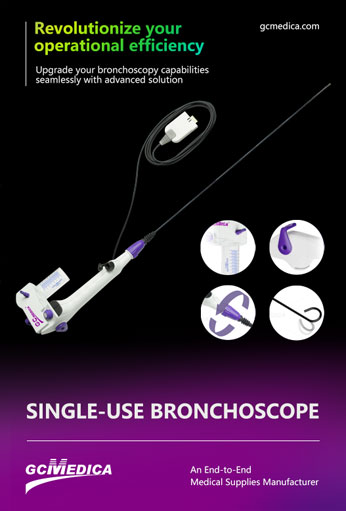
| Single Use Flexible Bronchoscopy > |


 Français
 Español
 Products
 About Us


