
 Français
 Español
 Products
 About Us


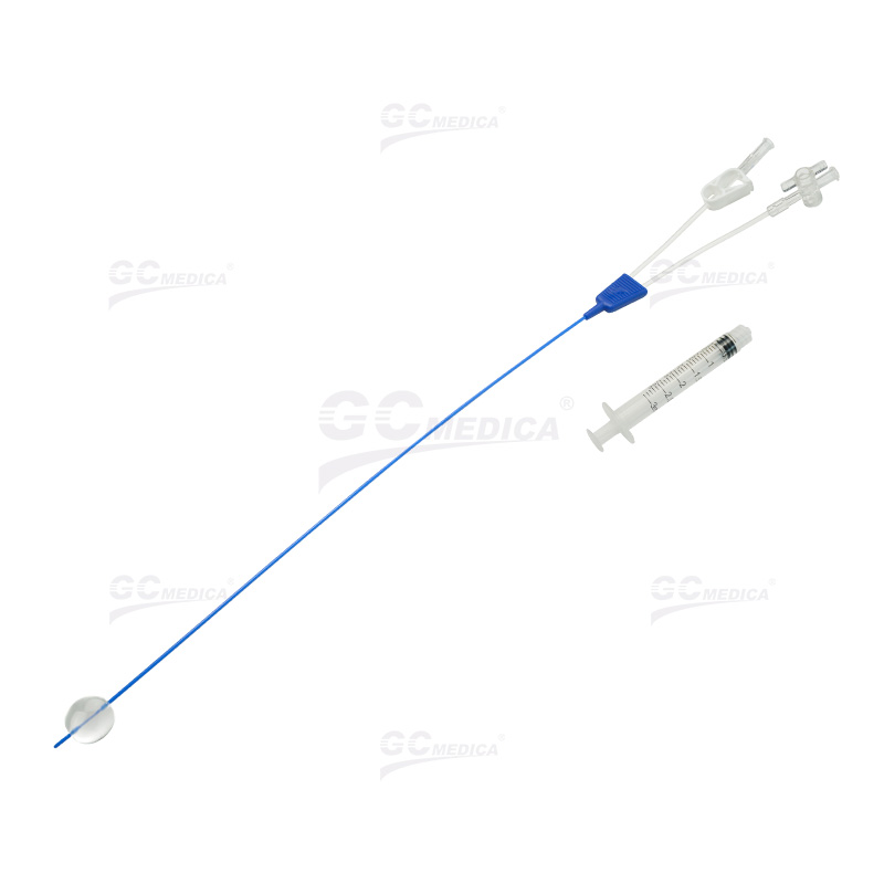
The HSG test balloon catheter is a specialized single‑use device designed for diagnostic imaging of the uterine cavity and fallopian tubes. By inflating its soft, atraumatic balloon within the uterus, contrast medium can be gently instilled under controlled pressure, allowing radiologists and gynecologists to visualize uterine shape, tubal patency, and potential intrauterine pathologies (e.g., adhesions, polyps). Its slim profile and malleable tip facilitate atraumatic cervical passage, while the radiopaque marker bands aid precise positioning under fluoroscopy.
Key features include an ultra‑smooth hydrophilic coating to minimize friction and patient discomfort, a low‑pressure compliant balloon that adapts to uterine contours, and a luer‑lock connector ensuring secure attachment of contrast syringes. The catheter shaft—available in multiple French sizes—combines rigidity for pushability with flexibility to follow the cervical canal’s curvature. A built‑in stop limits balloon inflation volume, reducing the risk of over‑distension.
| Specification | Description |
|---|---|
| Catheter French Size | 5 Fr, 7 Fr, or 9 Fr |
| Balloon Maximum Volume | 3–10 mL (compliant) |
| Inflation Pressure Range | 20–50 kPa |
| Total Catheter Length | 30 cm |
| Balloon Material | Medical‑grade silicone (late‑x free) |
| Shaft Material | Polyurethane core with hydrophilic coating |
| Radiopaque Markers | Dual marker bands at balloon shoulders |
| Connector | Standard luer‑lock |
| Sterilization | Ethylene oxide (ETO) |
| Packaging | Individually sealed, peel‑able pouch |
Constructed from biocompatible polyurethane and medical‑grade silicone, the HSG balloon catheter exhibits excellent kink resistance and chemical stability against contrast agents. The hydrophilic coating becomes slippery upon hydration, reducing insertion force and cervical trauma. Radiopaque markers, positioned immediately adjacent to the balloon’s proximal and distal ends, ensure accurate localization under X‑ray or fluoroscopic guidance.
During an HSG procedure, the patient is positioned supine on the radiology table and a small speculum is introduced for cervical visualization. After cleansing the cervix, the balloon catheter tip is gently advanced through the cervical os until the radiopaque markers lie within the uterine cavity. A small test inflation confirms proper placement and an adequate seal. Contrast medium is then injected under real‑time imaging: uniform filling of the uterine cavity followed by free spillage into the peritoneal cavity indicates patent tubes.
Advantages of this design include reduced discomfort, lowered risk of uterine perforation, and consistent balloon inflation pressures. The compliant silicone balloon adapts smoothly to various uterine anatomies, accommodating minor irregularities without excessive pressure. The hydrophilic coating eases passage through the cervix, particularly in nulliparous patients. Additionally, the luer‑lock connector prevents accidental disconnection during contrast injection.
In summary, the HSG test balloon catheter is an essential tool for fertility evaluation and uterine imaging. Its combination of atraumatic design, precise radiopaque positioning, and controlled inflation makes it a reliable choice for safe and effective hysterosalpingography. Whether assessing tubal patency or detecting uterine anomalies, this catheter offers clinicians the performance and patient comfort necessary for accurate diagnostic outcomes.
| HSG Test Balloon Catheter > |